输入基于细胞板多个域位置的图片数据
在CellProfiler(细胞分析器)中调整导出设置,并运行了传递途径后,你可以把数据导入到FCS Express中。FCS Express只需要关于实验设置的另外一项信息 。CellProfiler(细胞分析器)现在还不能以实验中用到的细胞板格式导出。一系列标准板格式定义可以在FCS Express网站下载。在本例中,我们将使用一个6孔细胞板模板,用户可在(Tutorial Sample Data archive)教程样本数据库中中的Multiple Field Location Plate Imaging (多个域位置的细胞板图像)文件夹中找到这个模板,其名称是Experiment.ini.细胞板格式定义文件必须被命名为Experiment.ini.,并且放置于您在CellProfiler(细胞分析器)中定义的默认输出文件夹中。
| 1. | 下载示例中的数据(Tutorial Sample Data archive)教程样本数据库中中的Multiple Field Location Plate Imaging (多个域位置的细胞盘图像)文件夹中下载。 |
| 2. | 下载MultipleFieldLocationPipelineCOMPLETED.cp传输途径到CellProfiler(细胞分析器)中。 |
| 3. | 请在CellProfiler(细胞分析器)中选择默认输入和输出文件夹,以便导入和存储数据,如图T24.56所示。 |
| 4. | 在cellProfiler(细胞分析器)中分析图片。 |
| 5. | 请确保Experiment.ini文件,位于(Tutorial Sample Data archive)教程样本数据库中中的Multiple Field Location Plate Imaging (多个域位置的细胞盘图像)文件夹下,该文件夹包含您输出的CellProfiler数据。 |
输入基于细胞板多个域位置的图片实验到FCS Express中
在开始以下步骤之前,我们先对FCS Express Preferences(FCS Express设置)做一点解释:
此File selection(文件选择)对话框必须激活:
| A. | 打开FCS Express User Options(FCS Express用户选项)选择文件→选项。 |
| B. | 选中When creating a new plot, always display the selection dialog(当创建一新绘图时,总是显示选择对话框 位于Files/Directories(文件/路径)选项。 |
(启用这个功能不是使用FCS Express的必需条件, 在本教程中选择这个功能只是为了方便)
| 6. | 打开FCS Express. |
| 7. | 打开FCS Express User Options(FCS Express用户选项)选择文件→选项 |
| 8. | 选择Cell Profiler Options(细胞分析工具选项)从Data Loading(数据导入)(图T24.61)。 |
| 9. | 选择Csv(分隔符)从delimiter(定界符)下拉菜单中。 |
| 10. | 选中Load Images(导入图片)复选框。 |
| 11. | 取消对复选框Load path names from Cell Profiler table of contents file(从内容文件的Cell Profiler(细胞分析器)表格中,导入路径名)复选框。 |
| 12. | 点击OK,来关闭FCS Express User Options(FCS Express用户选项)对话框。 |
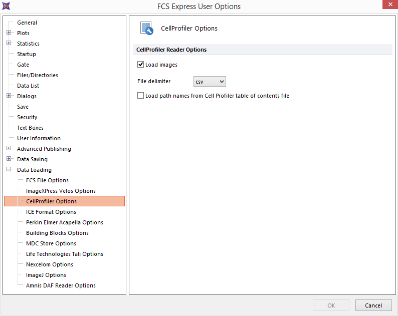
Figure T24.61 CellProfiler Data Loading Options for Importing Plate Based Multiple Field Location Imaging Data.
| 11. | 选择Insert(插入)→Other Plots(其它绘图)→Heat Map(热图)命令 (图T24.62)。 |

Figure T24.62 Choosing to Insert a Heat Map
| 12. | 请在版面上任意位置点击,Select a Data File(选择一数据文件)对话框将出现。 |
| 13. | 浏览默认输出文件夹你可以从CellProfiler(细胞分析器)中保存你的输出数据。 |
| 14. | 改变文件类型:下拉列表中到Cell Profiler (*.cpout)(图T24.63).现在,只会显示由CellProfiler(细胞分析器)输出的文档。在本例中,它们的名称分别为cells.cpout和nuclei.cpout。 |
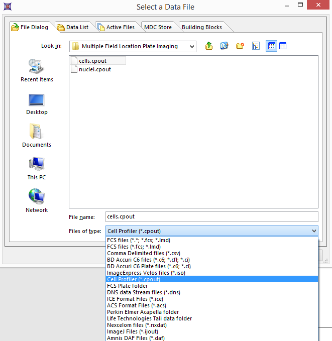
Figure T24.63 Choosing CellProfiler Files
| 15. | 高亮显示cells.cpout. |
| 16. | 点击Open(打开)。 |
注意:在本例中,我们将使用cells.cpout文件,该文件将显示数据,从对象分类到IdentifySecondaryObjects(确认次要对象)模块中的数据,这一模块对应于整个细胞面积。文件nuclei.cpout将显示CellProfiler中归为IdentifyPrimaryObjects(确定主要对象)模块中的数据,这个模块对应细胞的细胞核区域。
| 17. | 高亮Y-Axis Parameter(Y轴参数)你希望看到热度图,位于Open(打开) 2D Plots(二维绘图)对话框。对本例来说,我们选择Intensity_MeanIntensity_Cytoplasm(强度_平均强度_细胞质)(图T24.64)。 |
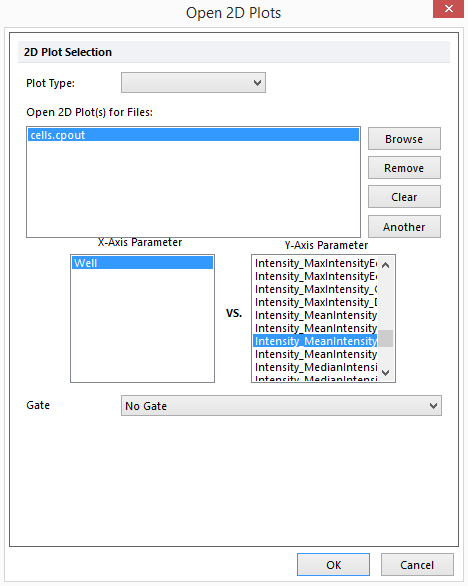
Figure T24.64 Opening a Heat Map and Choosing the Y-Axis Parameter from the Open 2D Plots Dialog
| 18. | 点击OK。 |
这时在版面上会出现一个热图,表明数据被成功导入(图T24.65)。
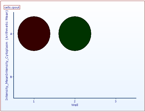
Figure T24.65 Example of a Heat Map Generated from a 6 Well Plate Based Multiple Field Location Imaging Data.
| 19. | 请右键点击Heat Map(热图)。 |
| 20. | 选择Create Gate(创建门)→Well(细胞孔)。 |
| 21. | 在孔A 01中建一个细胞孔门,命名为Gate 1(门1)。 |
| 22. | 请选择Insert Tab(插入标签)→2D Plots(二维绘图)→Picture Plot(图片绘图) |
| 23. | 在版面上的任何位置点击,来插入图片绘图。 |
| 24. | 从Select a Data File(选择一数据文件)对话框中选择cells.cpout文件。 |
| 25. | 点击Open(打开)。 |
| 26. | 选择Gate 1(门1)从Gate(门)下拉菜单中。 |
| 27. | 点击OK。 |
现在,图片绘图展示了来自A01细胞孔的4个域,就如微孔门Gate 1定义的一样(图T24.66)。
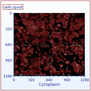
Figure T24.66 The picture plot has been gated on Gate 1, well A01, of the heat map. Four image fields may be observed in the image.
